English
  Français (French)
  Deutsch (German)
  Español (Spanish)
  Nederlands (Dutch)
  Italiano (Italian)
  Dansk/Norsk
  Svenska (Swedish)
  Polski (Polish)
  Čeština (Czech)
  Türkçe (Turkish)
  Русский (Russian)
  Arabic (اللغة العربية)
 Cell research made visible with
USB fluorescence microscopy
Dino-Lite helps researchers to form an image
Research into life-threatening diseases is of great importance. Miraculously a small striped fish with special light microscopy can play an important role. Professor Yung-Jen Chuang (47) from Taiwan is doing research with zebrafish using Dino-Lite fluorescence microscopes.
Within the National Tsing Hua University in Hsinchu, Taiwan, Professor Yung-Jen Chuang runs a laboratory for vascular biology. Vascular biology is the study of our circulatory system in all its forms, from the aorta to the smallest capillary in the brains. Professor Yung-Jen Chuang and his team are particularly interested in the molecular and cellular processes that occur when new blood vessels are formed from the existing blood vessels, a process that is called angiogenesis. The team is also investigating how tissue repair occurs after injury to vital organs such as heart or brains, and examines which reactions influence the blood circulation within a tumor. The studies also involve functional genomics that aims to identify what specific genes work harder, for instance to speed up regeneration. Obviously Professor Yung-Jen Chuang is leading a team that consists of a large number of researchers, an even greater number of zebrafish and Dino-Lite fluorescence microscopes.
Excitation and emission
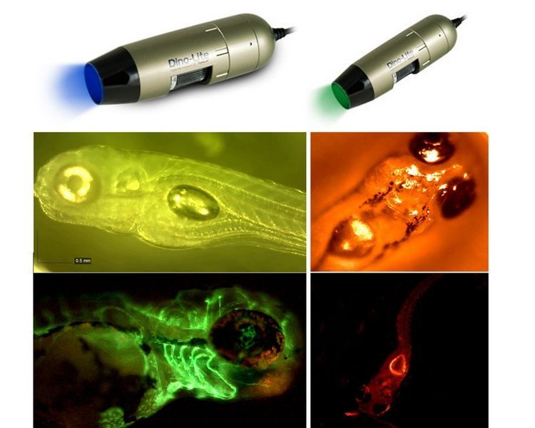
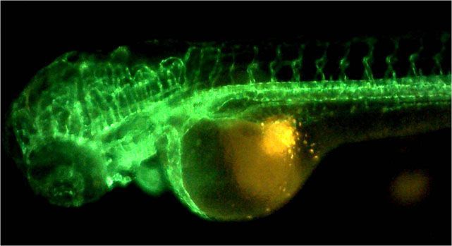
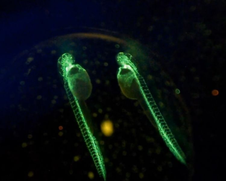
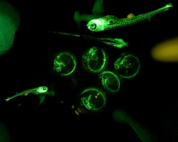
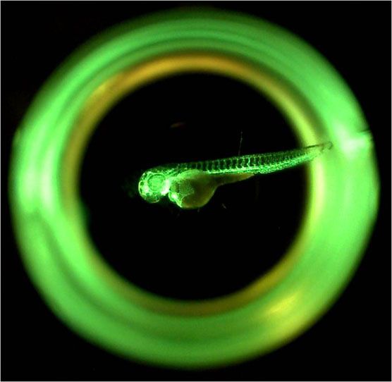
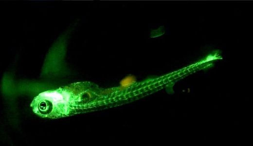
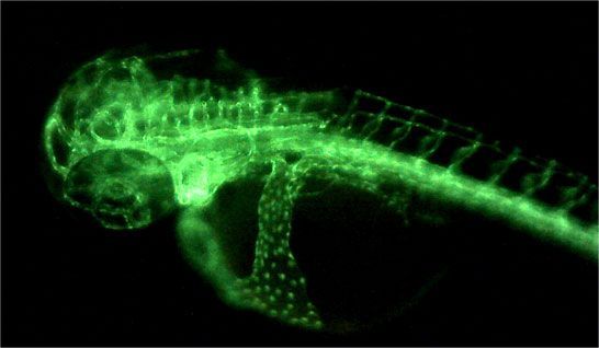
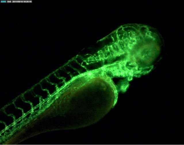
Dino-Lite has different fluorescence microscopes that are used by Professor Yung-Jen Chuang in his investigations. Fluorescence microscopy makes use of fluorescent dyes that give off light when it is irradiated with light of a shorter wavelength. This light, for example blue, is referred to as excitation light. The phosphors convert this light of a short wavelength into light of a longer wavelength, such as green or red. This emitted light is called emission light. The Dino-Lite fluorescence microscopes have a so-called excitation filter that limits the outgoing light to a certain wavelength (expressed in nanometers). In the same way, the emission light is going through a built-in emission filter that removes the excitation wavelength from the light bundle and the image only consists of the emission light. In this way, Professor Yung-Jen Chuang can see what happens in a living organism in real time. One of the animals he is using for this purpose is the zebrafish. Since the 90s of the last century many scientists are using zebrafish (scientific name Danio rerio) as a model organism. The genome (the total of genetic information in a cell) of a zebrafish is very similar to the genome of human beings. Zebrafish embryos are also translucent, making it possible to study various processes such as angiogenesis in an organism using fluorescence microscopy. The zebrafish is not naturally fluorescent, so how were scientists able to give the fish light?
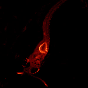
GFP, BFP, CFP, YFP and CherryFP
This became possible after Nobel laureate Osamu Shimomura discovered that the deep sea jellyfish Aequoria was naturally fluorescent and owes this property to a protein called green fluorescent protein (GFP). This protein can be compared to a beacon that brightened up biological research. In 1994 scientists were able to transfer this gene into the cells of higher organisms through transfection, the technique in which foreign DNA is placed into a cell. It was now possible to highlight each cell in color using fluorescence microscopy, which was a revolution in cell biology. But the scientists went further. By causing mutations in the GFP-gene, they were able to change the color of the fluorescence. Now there is not only green (GFP), but also blue (BFP), cyan (CFP), yellow (YFP) and red (CherryFP). Both the excitation and emission spectra of these variants differ so that different models Dino-Lite were made to observe the different colors. Most fluorescence microscopes have a traditional emission filter whereas Dino-Lite uses high-pass filters that provide enhanced imaging and improved sensitivity in a wider spectrum of the fluorescence.
Disease research
The GFP color variants give researchers the ability to let for instance arteries emit green light, immune cells red and bacteria blue. In this way an investigator can follow the occurrence and progression of a disease, such as cancer. In order to investigate the conduct of a cancer type, human cancer cells labelled with fluorescence are implanted into zebrafish embryos. This makes it possible to monitor all stages of the development of a cancer and allows research into the genes and substances involved in the development of cancer and the inhibition of the growth of the tumor cells. Professor Yung-Jen Chuang's team is obviously not the only one doing this type of research. Thousands of researchers all over the world are working with zebrafish and fluorescence microscopy to study diverse diseases like Parkinson's, multiple sclerosis and acute lymphocytic leukemia hoping to find solutions. In many cases, Dino-Lite literally helps researchers to form a good picture of the disease and healing processes that occur in an organism. With this knowledge drugs can be developed to inhibit, heal and even prevent diseases like cancer.
Affordable
Professor Yung-Jen Chuang worked with Dino-Lite to develop the fluorescence digital microscopes: "I am delighted that the Dino-Lite fluorescence microscopes are of good quality and affordable. Moreover, they are easy to use. Thus, we can enable more researchers to work after minimal training, and also enlist various sets of Dino-Lites that we have for educational purposes. It is easy to show the images on a laptop, and we can store both video and still images to study changes in tissue better." Currently, Yung-Jen Chuang has achieved most successes in studying the interaction between a host such as a human and a fungal pathogen such as Candida albicans.
What Dino-Lite models does Professor Yung-Jen Chuang use?
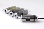
In his laboratory in Taiwan Professor Yung-Jen Chuang uses three types of fluorescence microscope and a general Dino-Lite. The Dino-Lite fluorescence microscopes are the smallest fluorescence microscopes in the world. The user can switch from colored LEDs to the built-in white LED, which is useful for focusing and locating the object.
Dino-Lite AM4115T-GFBW
The Dino-Lite AM4115T-GFBW is a fluorescence microscope equipped with blue LEDs. The emission filter of 510 nanometers ensures that Green Fluorescent Protein (GFP) shows up.
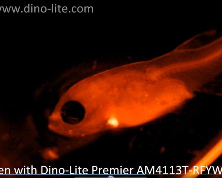
Dino-Lite AM4115T-RFYW
The Dino-Lite AM4115T-RFYW uses yellow LEDs and an emission filter of 610 nm. This microscope is very useful in developmental biology, pathology and anatomy.
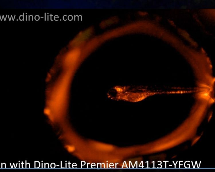
Dino-Lite AM4115T-YFGW
The Dino-Lite AM4115T-YFGW features green LEDs for excitation. The 570 nm emission filter of this fluorescence microscope ensures that orange to red fluorescence such as RFP (Red Fluorescent Protein) shows up.
Dino-Lite AM4113ZT
AM4113ZT is not a fluorescence microscope, but is also used in the lab of Professor Yung-Jen Chuang. This is one of the top selling Dino-Lite models. This USB microscope is provided with a rotatable polarizing filter so that research on shiny objects becomes simpler, because disturbing reflections are reduced significantly.
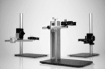
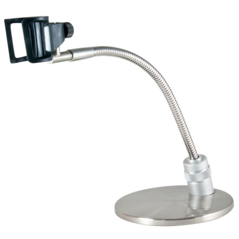 Round metal base with flexible arm and holder for the Dino-Lite. The MS33W has the possiblility to vary the height (appr. 15 mm) with a turning knob in the base.
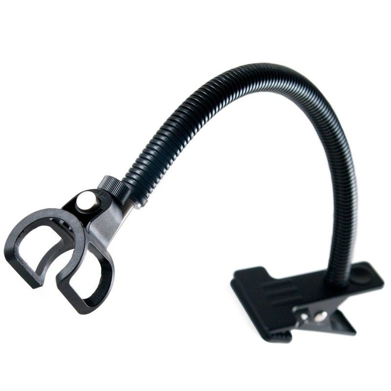 The MS23B is a desktop clamp with flexible gooseneck that can be clamped to a desktop or any other surface of up to 3cm thick.
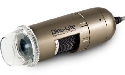 By using a special polarization filter, the AM4113ZT is the right choice when working with high glare materials such as plastics and metals.
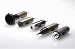
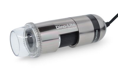 The Dino-Lite AM7013MZT has a 5 megapixel sensor for crystal clear images, even under higher magnification. The built-in adjustable polarizer reduces glare and reflection on shiny objects.
 By using a special polarization filter, the AM4013MZTL is the right choice when working with high glare materials such as plastics and metals. This model features an aluminum alloy housing for enhanced protection and durability and the best look and feel.
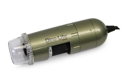 By using a special polarization filter, the AM4113ZTL is the right choice when working with high glare materials such as plastics and metals.
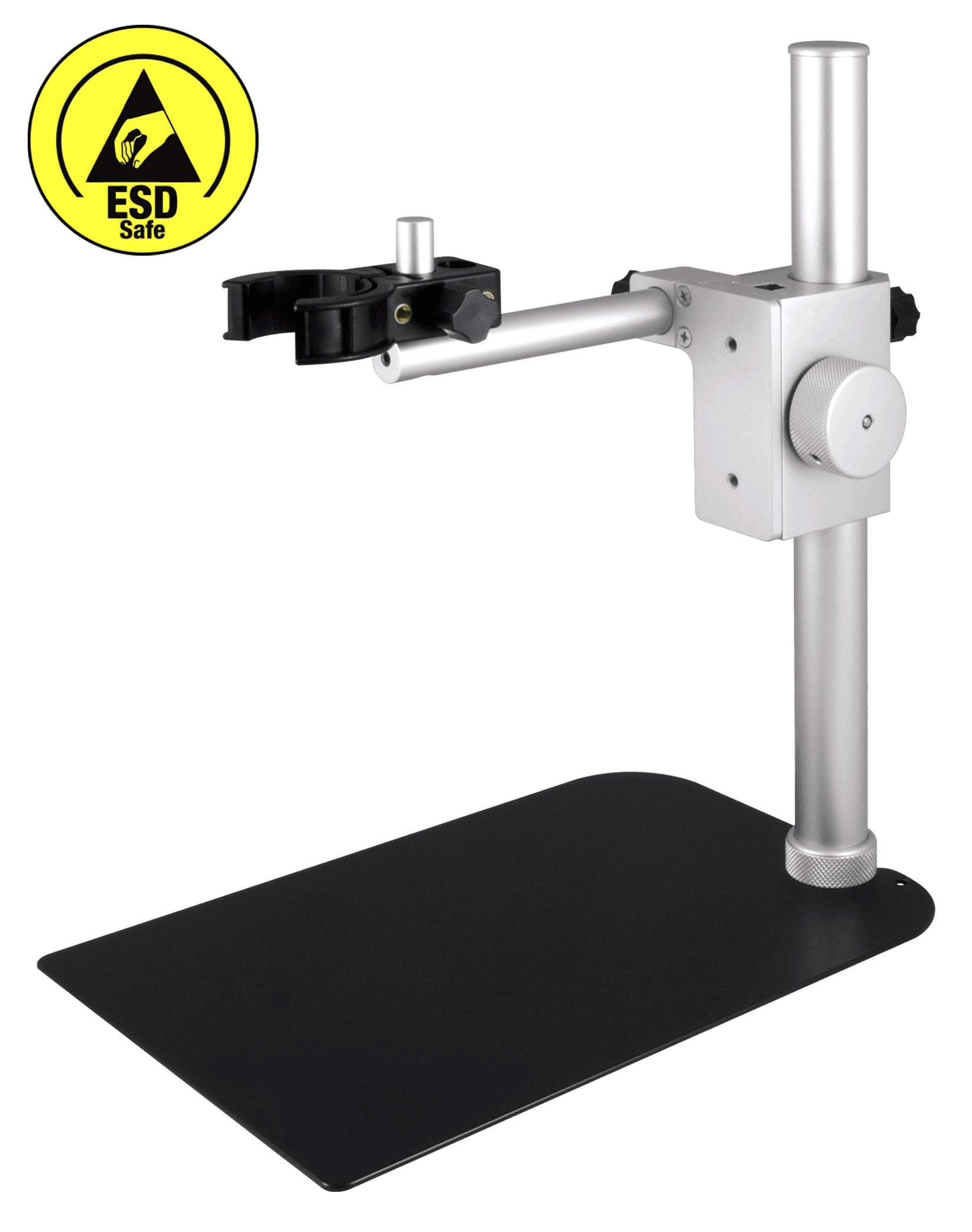 The Dino-Lite RK-06-AE stand is a sturdy and stable mid-range stand solution that can be used with all Dino-Lite digital microscopes. It is constructed of stainless steel and lightweight aluminum and offers precise fine-focus adjustment as well as a quick vertical release function.
 The Dino-Lite AD7013MZT has a 5 megapixel sensor for crystal clear images, even under higher magnification. The built-in polarizer reduces the shiny effect on reflecting materials, such as metals, plastics, jewelry, electronics etc.