Dansk/Norsk
  English
  Français (French)
  Deutsch (German)
  Español (Spanish)
  Nederlands (Dutch)
  Italiano (Italian)
  Svenska (Swedish)
  Polski (Polish)
  Čeština (Czech)
  Türkçe (Turkish)
  Русский (Russian)
  Arabic (اللغة العربية)
 Fluorescence Guided Surgery, FGS.
For surgeons it is of the utmost importance to be able to able to see accurately the tumour margins during the surgery. This is of particular importance for resection of metastatic diseases. An important and For FGS, clinically approved fluorescent dyes are being used to label the abnormal cells or diseased tissue. Illuminating the area of interest with the corresponding excitation light and the use of a correct emission filter in front of the imaging device, the cancerous tissue will light up and can be removed easily and accurately. With this method smaller tumours can be removed, opposed to the standard methods of visualisation and detection. FGS is nowadays considered as an effective way to carry out cancer surgery in comparison to the conventional surgery. Researchers around the world are exploring the possibilities of this emerging technology.
In the following 4 open access research papers, several different Dino-Lite Fluorescence microscopes have been used to perform Fluorescence Guided Surgery successfully.
-
FGS of Liver Metastasis in Orthotopic Nude-Mouse Models:
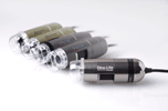
The fluorescence Dino-Lite model AM4113T-GFBW has been used to visualize the green fluorescence protein (GFP) labelled tumour before, during and after the surgery.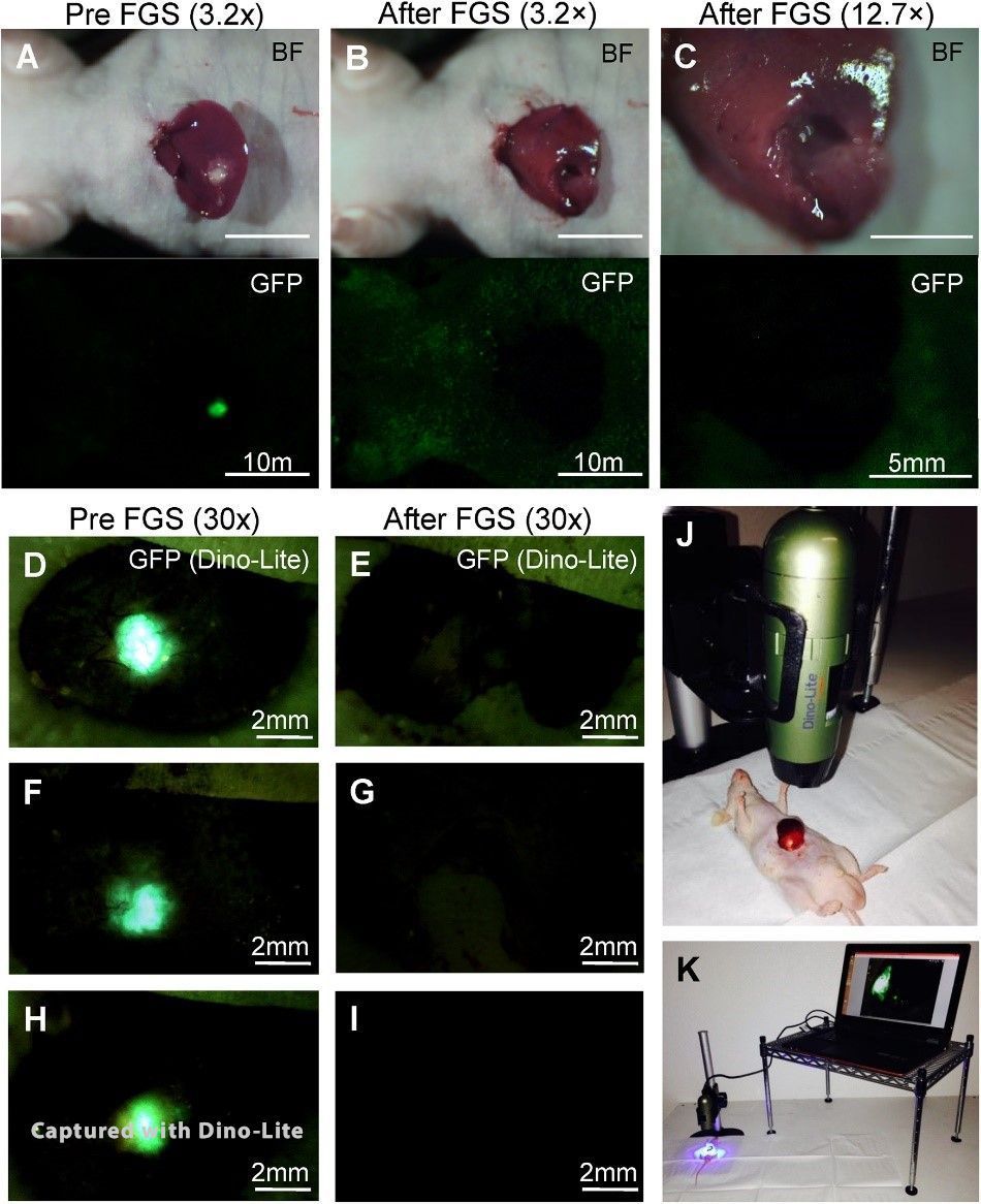
The researchers mentioned below have performed a trial to show prove that FGS is an effective method for liver metastasis. Fourteen 14 mice of the created orthotopic liver metastasis model had been randomly divided in 2 groups. One group was treated with BLS (Bright Light Surgery) and the other group was treated with FGS by using the Dino-Lite fluorescence digital microscope. Post-surgical residual was clearly present with the BLS treated mice, while the FGS group showed no sign of residual tumor.
Paper: Murakami T, Hiroshima Y, Zhang Y, Chishima T, Tanaka K, Bouvet M, et al (2015) Fluorescence-Guided Surgery of Liver Metastasis in Orthotopic Nude-Mouse Models.
Link: PloS ONE 10(10): e0138752. Doi:10.1371/journal.pone.0138752
-
FGS using In Situ GFP Labeling with a Telomerase-Dependent Adenovirus in an Orthotopic Mouse Model
In this research article they demonstrate that labelling colon-cancer liver metastasis in-situ with the GFP OBP-401 adenovirus is a powerful tool to complete resection with FGS. Compared to the conventional BLS ( Bright Light Surgery) it reduced the recurrence rate and prolonged the over-all survival. FGS was performed under GFP guidance using amongst others the Dino-Lite fluorescence microscope AM4113T-GFBW.
Paper: Yano S, Takehara K, Miwa S, Kishimoto H, Hiroshima Y, Murakami T, et al. (2016) Improved Resection and Outcome of Colon-Cancer Liver Metastasis with Fluorescence-Guided Surgery Using In Situ GFP Labeling with a Telomerase-Dependent Adenovirus in an Orthotopic Mouse Model.
Link: PLoS ONE 11(2): e0148760. doi:10.1371/journal. pone.0148760 -
Color-coded FGS to Resect the Tumor along with the Tumor Microenvironment
“Color coded FGS is an effective method to completely resect cancer cells along with the stromal cells in the TME which interact in a highly-complex pattern.“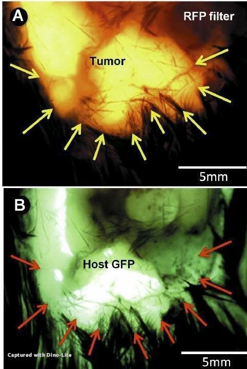
Results: Using the Dino-Lite, subcutaneous tumors and the tumor microenvironment were clearly visualized and resected.” During this research they have used the AM4113T-YFGW to visualize the EL-4-RFP stained cells and the AM4113T-GFBW for the GFP stained cells.Paper: Color-coded Imaging Enables Fluorescence-Guided Surgery to Resect the Tumor Along with the Tumor Microenvironment in a Syngeneic Mouse Model of EL-4 Lymphoma. Hasegawa K, Suetsugu A, Nakamura M, Matsumoto T, Kunisada T, Shimizu M, Saji S, Moriwaki H, Bouvet M, and Hoffman R.
Link: Anticancer research 36: 4443-4448 (2016). doi:10.21873/anticanres.10988
-
Color-coding cancer and stromal cells with genetic reporters enhances FGS
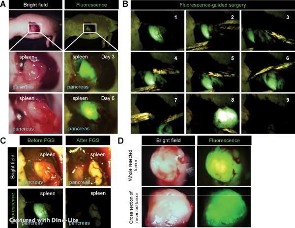 Also here GFP containing the OBP401 was used to label the cancer cells of the pancreatic cancer PDOX. The PDOX was previously grown in a RFP transgenic mouse. This dual color-coding enabled FGS to completely resect the pancreatic tumors including stroma. “Dual-colored FGS significantly prevented local recurrence, while bright-light surgery (BLS) or single color could not”. “The Dino-Lite mobile imaging system was used for imaging in live mice.” “This all-in-one compact digital camera makes the Dino-Lite imaging system easily transportable and thereby suitable for FGS”.
Paper: S Yano, Y Hiroshima, A Maawy, H Kishimoto, A Suetsugu, S Miwa, M Toneri, M Yamamoto, M H G Katz, J B Fleming, Y Urata, H Tazawa, S Kagawa, M Bouvet, T Fujiwara and R M Hoffman. Color-coding cancer and stromal cells with genetic reporters in a patient-derived orthotopic xenograft (PDOX) model of pancreatic cancer enhances fluorescence-guided surgery.
Link: Cancer Gene Therapy 22, 344-350 (July 2015) | doi:10.1038/cgt.2015.26
 Dino-Lite AD7013MZT har en 5 megapixel sensor der giver krystalklare billeder, selv under større forstørrelse. Den indbyggede polarisator reducerer den skinnende effekt på reflekterende materialer såsom metaller, plastik, smykker, elektronik osv.
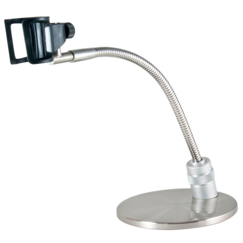 Rund metalbase med fleksibel arm og holder til Dino-Lite. MS33W har mulighed for at variere højden (ca. 15 mm) med en drejeknap i basen.
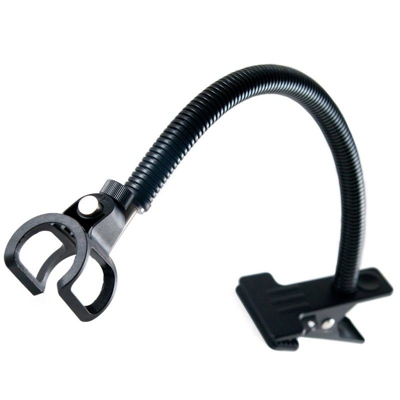 MS23B er en skrivebordsklemme med fleksibel svanehals, som kan fastspændes på et skrivebord eller enhver anden overflade med op til 3 centimeters tykkelse.
 Ved hjælp af et specielt polarisationsfilter, er AM4113ZT det rigtige valg til arbejde med materialer med høj blænding såsom plastik og metaller.
 Dino-Lite AM7013MZT har en 5 megapixel sensor der giver krystalklare billeder, selv under større forstørrelse. Den indbyggede justerbare polarisator reducerer blænding og refleksion på skinnende objekter.
 Ved hjælp af et specielt polarisationsfilter, er AM4013MZTL det rigtige valg til arbejde med materialer med høj blænding såsom plastik og metaller. Denne model har et aluminiumlegeringshus til bedre beskyttelse og holdbarhed og har det bedste udseende.
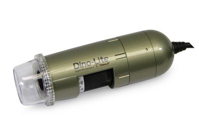 Ved hjælp af et specielt polarisationsfilter, er AM4113ZTL det rigtige valg til arbejde med materialer med høj blænding såsom plastik og metaller.
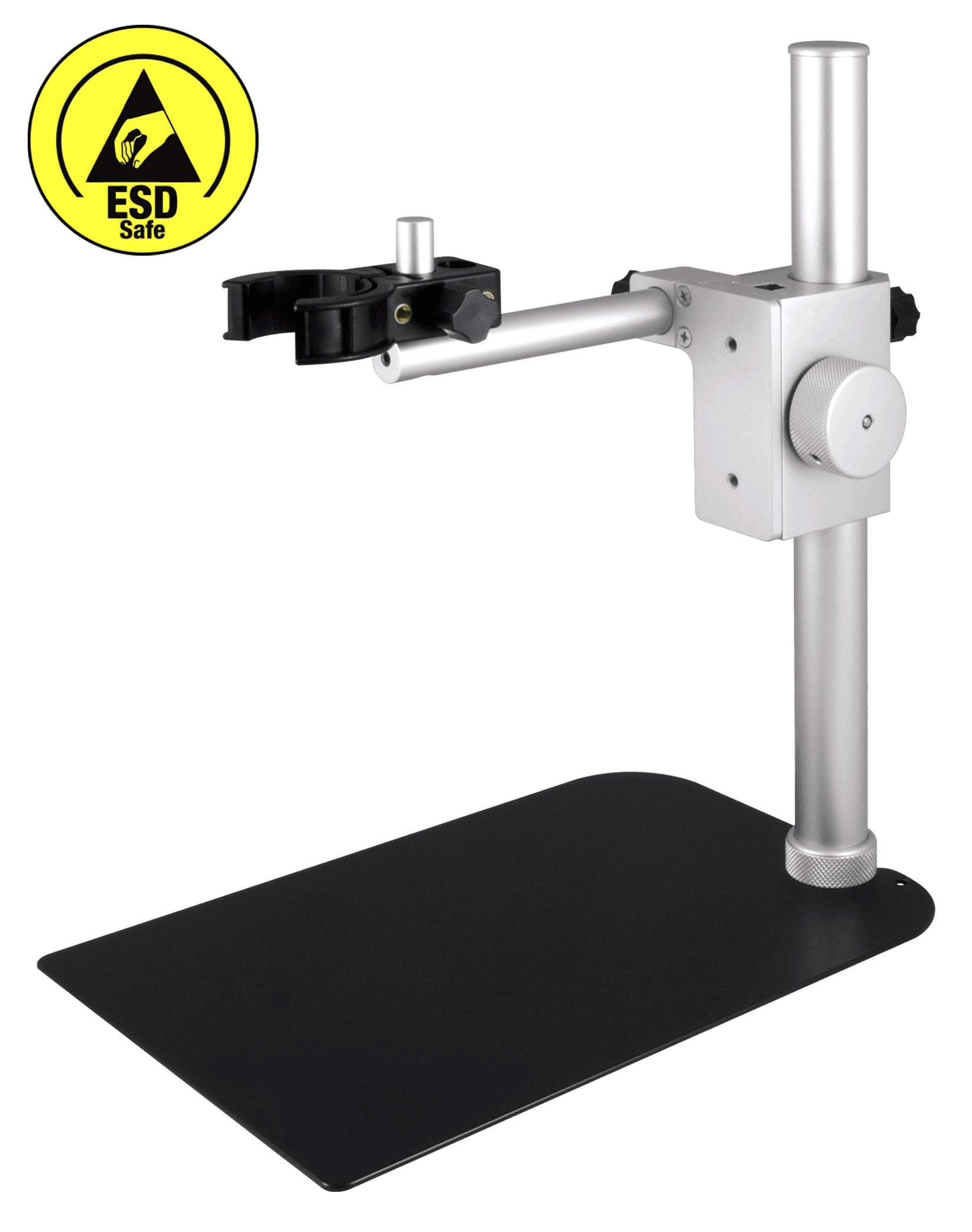 Dino-Lite RK-06-AE understøtning er et robust og stabilt produkt til understøtning i mellemklassen, der kan bruges sammen med alle Dino-Lite digitale mikroskoper. Den er konstrueret af rustfrit stål og letvægtsaluminium og har præcis finfokusjustering samt en hurtig lodret frigivelsesfunktion.
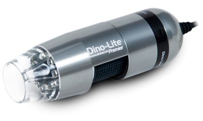 Dino-Lite professionel model med aluminiumlegeringshus (med MicroTouch). Aluminiumshuset giver bedre beskyttelse og holdbarhed og har det bedste udseende.